M
Phase (Mitosis Phase)
This is the most
dramatic period of the cell cycle, involving a major reorganisation of
virtually all components of the cell. Since the number of chromosomes in
the parent and progeny cells is the same, it is also called as equational
division.
Though for convenience
mitosis has been divided into four stages of nuclear division, it is essential to understand that cell division
is a progressive process and very clear-cut lines cannot be drawn between
various stages.
Mitosis is divided
into the following four stages:
·
Prophase
·
Metaphase
·
Anaphase
·
Telophase
Prophase:
Prophase which is the
first stage of mitosis follows the S and G2 phases
of interphase. In the S and G2 phases, the new DNA molecules formed are not distinct but
interwined. Prophase is marked by the initiation of condensation of
chromosomal material. The chromosomal material becomes untangled during the
process of chromatin condensation (below figure).
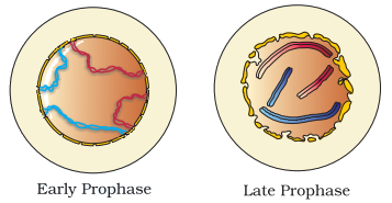
The
centriole, which had undergone duplication during S phase of interphase, now
begins to move towards opposite poles of the cell.
The completion of
prophase can thus be marked by the following characteristic events:
·
Chromosomal
material condenses to form compact mitotic chromosomes. Chromosomes are seen
to be composed of two chromatids attached together at the centromere.
·
Initiation
of the assembly of the mitotic spindle,
the microtubules, the proteinaceous components of the cell cytoplasm help
in the process.
Cells at the end of prophase,
when viewed under the microscope, do not show golgi complexes,
endoplasmic reticulum, nucleolus and the nuclear envelope.
Metaphase:
The complete
disintegration of the nuclear envelope marks the start of the second phase
of mitosis, hence the chromosomes are spread through the cytoplasm of the
cell.
By this stage,
condensation of chromosomes is completed and they can be observed clearly
under the microscope.
At this stage,
metaphase chromosome is made up of two sister chromatids, which are held
together by the centromere (below figure).
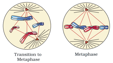
Small
disc-shaped structures at the surface of the centromeres are
called kinetochores. These structures serve as the sites of
attachment of spindle fibres (formed by the spindle fibres) to the
chromosomes that are moved into position at the centre of the cell.
Hence, the metaphase
is characterised by all the chromosomes coming to lie at the equator with
one chromatid of each chromosome connected by its kinetochore to
spindle fibres from one pole and its sister chromatid connected
by its kinetochore to spindle fibres from the opposite pole (above
figure).
The plane of alignment
of the chromosomes at metaphase is referred to as the metaphase plate.
The key features
of metaphase are:
·
Spindle
fibres attach to kinetochores of chromosomes.
·
Chromosomes
are moved to spindle equator and get aligned along metaphase plate through
spindle fibres to both poles.
Anaphase:
At the onset of
anaphase, each chromosome arranged at the metaphase plate is split
simultaneously and the two daughter chromatids, now referred to as
chromosomes of the future daughter nuclei, begin their migration
towards the two opposite poles.
As each chromosome
moves away from the equatorial plate, the centromere of
each chromosome is towards the pole and hence at the leading edge,
with the arms of the chromosome trailing behind (below figure).
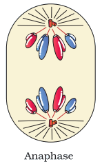
Thus, anaphase stage
is characterised by the following key events:
·
Centromeres
split and chromatids separate.
·
Chromatids
move to opposite poles.
Telophase:
At the beginning of
the final stage of mitosis, i.e., telophase, the chromosomes that have
reached their respective poles decondense and lose their individuality.
The
individual chromosomes can no longer be seen and chromatin
material tends to collect in a mass in the two poles (below figure).
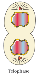
This is the stage
which shows the following key events:
·
Chromosomes
cluster at opposite spindle poles and their identity is lost as discrete
elements.
·
Nuclear
envelope assembles around the chromosome clusters.
·
Nucleolus,
golgi complex and ER reform.
Cytokinesis:
Mitosis accomplishes
not only the segregation of duplicated chromosomes into daughter nuclei
(karyokinesis), but the cell itself is divided into two daughter cells by
a separate process called cytokinesis at the end of which cell division
is complete (below figure).
In an animal cell,
this is achieved by the appearance of a furrow in the plasma
membrane. The furrow gradually deepens and ultimately joins in
the centre dividing the cell cytoplasm into two.
Plant cells, however, are enclosed by a relatively
inextensible cell wall, thererfore they undergo cytokinesis by a
different mechanism.
In plant cells, wall
formation starts in the centre of the cell and grows outward to meet the
existing lateral walls.
The formation of the
new cell wall begins with the formation of a simple precursor, called the
cell-plate that represents the middle lamella between the walls of
two adjacent cells.
At the time of
cytoplasmic division, organelles like mitochondria and plastids get
distributed between the two daughter cells.
In some organisms, karyokinesis is not followed by
cytokinesis as a result of which multinucleate condition arises leading to the
formation of syncytium (e.g., liquid endosperm in coconut).
The significance of Mitosis:
·
Mitosis
or the equational division is usually restricted to the diploid cells only. However,
in some lower plants and in some social insects haploid cells also divide by
mitosis.
·
Mitosis
results in the production of diploid daughter cells with identical genetic
complement usually.
·
The
growth of multicellular organisms is due to mitosis. Cell growth results in
disturbing the ratio between the nucleus and the cytoplasm.
·
It, therefore,
becomes essential for the cell to divide to restore the nucleo-cytoplasmic
ratio. A very significant contribution of mitosis is cell repair.
·
The
cells of the upper layer of the epidermis, cells of the lining of the gut, and
blood cells are being constantly replaced.
·
Mitotic
divisions in the meristematic tissues – the apical and the lateral cambium,
result in a continuous growth of plants throughout their life.
|
|
|
|