Anatomy of Dicotyledonous
and Monocotyledonous Plants

Dicotyledonous Root:

T.S.:
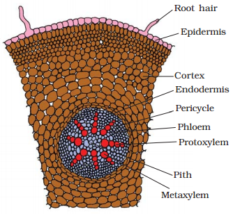
Dicot root (Primary)
The above figure shows the transverse section of the
sunflower root. The internal tissue organisation is as follows:
• Its outermost layer is epidermis. Many of the epidermal
cells protrude in the form of unicellular root hairs.
• The cortex consists of several layers of thin-walled
parenchyma cells with intercellular spaces and the innermost layer of the
cortex is called endodermis.
• It comprises a single layer of barrel-shaped cells without
any intercellular spaces. The tangential as well as radial walls of the
endodermal cells have a deposition of water impermeable, waxy material-suber in-in the form of casparian
strips.

• Next to endodermis lies a few layers of thick-walled parenchyomatous cells referred to as pericycle.
• Initiation of lateral roots and vascular cambium during the
secondary growth takes place in these cells. The pith is small or
inconspicuous. The parenchymatous cells which lie
between the xylem and the phloem are called conjuctive
tissue.
• There are usually two to four xylem and phloem patches.
Later, a cambium ring develops between the xylem and phloem. All tissues on the
inner side of the endodermis such as pericycle,
vascular bundles and pith constitute the stele.

Monocotyledonous Root:
T.S. : Monocot root
• The anatomy of the monocot root is similar to the dicot
root in many respects.
• It has epidermis, cortex, endodermis, pericycle,
vascular bundles and pith.
• As compared to the dicot root which have fewer xylem
bundles, there are usually more than six (polyarch)
xylem bundles in the monocot root.
• Pith is large and well developed.
• Monocotyledonous roots do not undergo any secondary growth.

Dicotyledonous Stem:

T.S. of stem : Dicot
• The dicotyledonous stem has the epidermis is the outermost
protective layer of the stem, covered with a thin layer of cuticle, it may bear
trichomes and a few stomata.
• The cells arranged in multiple layers between epidermis and
pericycle constitutes the cortex.
• It consists of three sub-zones. The outer hypodermis,
consists of a few layers of collenchymatous cells just
below the epidermis, which provide mechanical strength to the young stem.
Cortical layers below hypodermis consist of rounded thin walled parenchymatous cells with conspicuous intercellular
spaces.
• The innermost layer of the cortex is called the endodermis.
The cells of the endodermis are rich in starch grains and the layer is also
referred to as the starch sheath. Pericycle is
present on the inner side of the endodermis and above the phloem in the form of
semi-lunar patches of sclerenchyma.

• In between the vascular bundles there are a few layers of
radially placed parenchymatous cells, which
constitute medullary rays. A large number of vascular bundles are arranged in a
ring which is a characteristic of dicot stem. Each vascular bundle is conjoint,
open, and with endarch protoxylem.
A large number of rounded, parenchymatous cells with
large intercellular spaces occupying the central portion of the stem constitute
the pith.

Monocotyledonous Stem:

T.S. of stem: Monocot
• The monocot stem has a sclerenchymatous
hypodermis, a large number of scattered vascular bundles, each surrounded by a sclerenchymatous bundle sheath, and a large, conspicuous parenchymatous ground tissue.
• Vascular bundles are conjoint and closed.
• Peripheral vascular bundles are generally smaller than the
centrally located ones.
• The phloem parenchyma is not available, and
water-containing cavities are present within the vascular bundles.

Dorsiventral (Dicotyledonous) Leaf:

T.S. of leaf : Dicot
The dorsiventral leaf through the
lamina shows three main parts, namely,
·
Epidermis
·
Mesophyll
·
Vascular system
• Conspicuous cuticle are available in the epidermis which
covers both the upper surface (adaxial epidermis) and
lower surface (abaxial epidermis) of the leaf. The abaxial epidermis generally bears more stomata and adaxial epidermis, may even lack stomata. The tissue
between the upper and the lower epidermis is called the mesophyll.

• Mesophyll, which has chloroplasts, carry out photosynthesis
and is made up of parenchyma. It has two types of cells – the palisade
parenchyma and the spongy parenchyma.
• The adaxially placed palisade
parenchyma is made up of elongated cells, which are arranged vertically and
parallel to each other. The oval or round and loosely arranged spongy
parenchyma is located below the palisade cells and spreads to the lower
epidermis. Large spaces and air cavities are available between these
cells.
• Vascular system includes vascular bundles, as in the veins
and the midrib and the sizes of the vascular bundles are dependent on the size
of the veins. The veins vary in thickness in the reticulate venation of the
dicot leaves. The vascular bundles are encircled by a layer of thick walled
bundle sheath cells.

Isobilateral (Monocotyledonous) Leaf:

T.S. of leaf: Monocot
• In an isobilateral leaf, the stomata are on both the
surfaces of the epidermis and the mesophyll is not distinguished into palisade
and spongy parenchyma.
• In grasses, certain adaxial epidermal
cells along the veins modify themselves into large, empty, colourless cells,
called bulliform cells.
• When the bulliform cells in the
leaves have absorbed water and are turgid, the leaf surface is exposed. When
they are flaccid due to water stress, they make the leaves curl inwards to
minimise water loss.
• The parallel venation in monocot leaves is reflected in the
near similar sizes of vascular bundles (except in main veins).


|
|
|
|